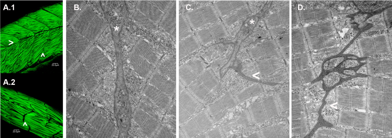
Fig. 6 Obscurin depletion disrupts somite architecture. A: Embryos injected with MO2 (6 ng; A.2) or a control morpholino CMO2 (6 ng; A.1) were fixed and immunostained for α-actinin at 72 hpf. Note that in the morphant embryos, there are no detectable horizontal myoseptae (>) and only rudimentary transverse myoseptae (∧) compared to control embryos. Elongated, disarrayed myofibrils often extend beyond the length of a normal somite. B-D: Electron micrographs of transverse myoseptae from control [6 ng CMO2 (B)] and obscurin morphant [3 ng (C) and 6 ng (D) of MO2] embryos. Morphant embryos displayed rudimentary transverse myoseptae (C:*, C,D: <) at the ends of the skeletal myocytes compared to the well-organized transverse myoseptae of control embryos (B: *).
Image
Figure Caption
Figure Data
Acknowledgments
This image is the copyrighted work of the attributed author or publisher, and
ZFIN has permission only to display this image to its users.
Additional permissions should be obtained from the applicable author or publisher of the image.
Full text @ Dev. Dyn.

