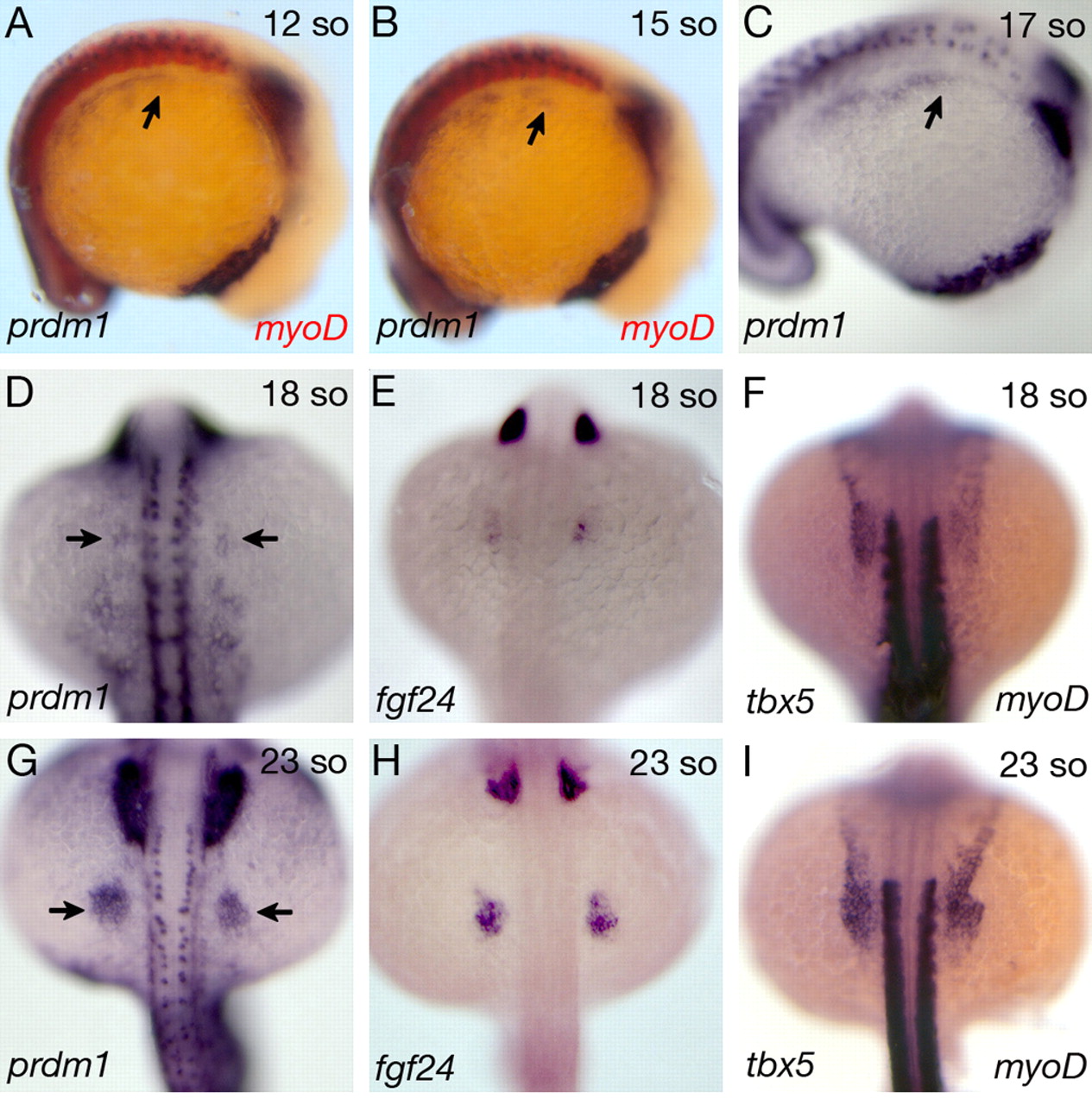
Fig. 2 Expression of prdm1 compared with fgf24 and tbx5 during limb bud initiation. (A-C) prdm1 whole-mount in situ hybridization at embryonic stages prior to limb bud initiation. Lateral views of a 12-somite (A) and a 15-somite (B) stage embryo revealing prdm1 (blue) and myod (red) expression. Note prdm1 expression overlapping with myod in the somites. (C) Lateral view of a 17-somite stage embryo. Arrows in A-C reveal the most anterior limit of prdm1 expression within the lateral plate mesoderm. (D-F) Dorsal views of 18-somite stage embryos hybridized with prdm1 (D), fgf24 (E) or tbx5+myod (F) riboprobes. Arrows in D point towards the onset of prdm1 expression in the pectoral fin primordia. Note that the prdm1 and fgf24 expression domains are very similar, and that the tbx5 expression domain is broader than the prdm1 domain. (G-I) Dorsal views of 23-somite stage embryos hybridized with prdm1 (G), fgf24 (H) or tbx5+myod (I) riboprobes. Arrows in G point towards the expanded prdm1 expression domain in the pectoral fin.
Image
Figure Caption
Figure Data
Acknowledgments
This image is the copyrighted work of the attributed author or publisher, and
ZFIN has permission only to display this image to its users.
Additional permissions should be obtained from the applicable author or publisher of the image.
Full text @ Development

