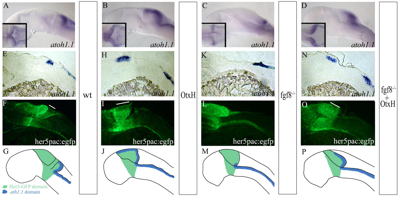
Fig. 4 Formation of the granule cell population in fgf8-/-; OtxH embryos. atoh1a expression in wild-type (A), OtxH (B), fgf8-/- (C) and fgf8-/-; OtxH (D) whole-mount embryos. MyoD expression in the somite was concomitantly detected to confirm fgf8-/-genotype. (E-O) Para-saggital 10 μm sections, anterior towards the left, of wild-type her5pac:egfp (E,F), OtxH (H,I), fgf8-/- (K,L) and fgf8-/-; OtxH (N,O) brains, showing co-localisation of atoh1a (E,H,K,N) and GFP expression (F,I,L,O). Granule cells absent in fgf8-/- are rescued in fgf8-/-; OtxH and are GFP positive (white lines on fluorescent pictures represent the length of the I>atoh1a domain in the upper rhombic lip obtained from bright field pictures). (G,J,M,P) Cartoons summarising GFP (in green) and atoh1a (in blue) expressions in wild-type (G), OtxH (J), fgf8-/- (M) and fgf8-/-; OtxH (P) embryos carrying the transgene her5pac:egfp.
Image
Figure Caption
Figure Data
Acknowledgments
This image is the copyrighted work of the attributed author or publisher, and
ZFIN has permission only to display this image to its users.
Additional permissions should be obtained from the applicable author or publisher of the image.
Full text @ Development

