
Image
|
Figure Caption
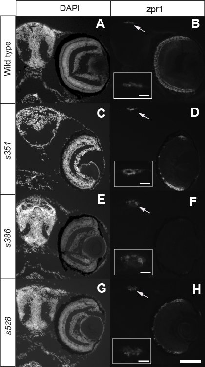
Fig. S1 Pineal Photoreceptors Are Present in Retinal Photoreceptor Degeneration Mutants Coronal sections of the forebrain at 7 dpf were stained with DAPI (A, C, E, and G) and zpr1, a marker of both retinal and pineal photoreceptors (B, D, F, and H). Pineal photoreceptors (arrow and inset) were consistently present in mutants in which retinal photoreceptors were depleted (D, F, and H). Scale bar is 100 μm for A-J and 25 μm for the insets.
Figure Data
Acknowledgments
This image is the copyrighted work of the attributed author or publisher, and
ZFIN has permission only to display this image to its users.
Additional permissions should be obtained from the applicable author or publisher of the image.
Full text @ PLoS Genet.