
|
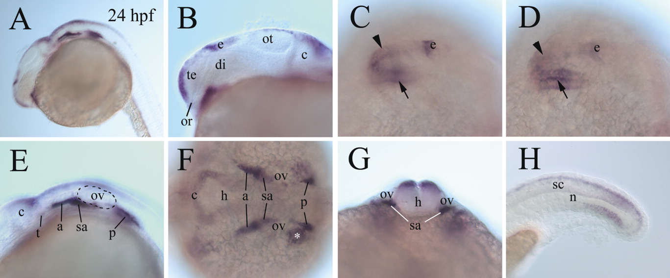
Fig. 3 Expression of cadherin-6 message in 24 hours postfertilization (hpf) embryos. A,B: B is a higher magnification of the fore- and midbrain regions of the embryo in A, with anterior to the left and dorsal up. C,D: Higher magnification dorsolateral views (anterior to the left) of the forebrain region of the same whole-mount embryo, with C focusing on cadherin-6 expression in the dorsal forebrain (arrowhead) and D focusing on cadherin-6 expression in the ventral diencephalon (arrow). E: A higher magnification of the hindbrain region of the embryo in A (anterior to the left and dorsal up). F: A higher magnification dorsal view (anterior to the left) of the hindbrain region of a whole-mount embryo. The asterisk indicates cadherin-6 expression outside the nervous tissue. G: A frontal view of the hindbrain region (dorsal up) at the level of otic vesicle (ov), of a whole-mount embryo. H: A higher magnification lateral view (anterior to the left and dorsal up) of a whole-mount tail region. c, cerebellum; sa, statoacoustic ganglion. The remaining abbreviations are the same as in Figures 1 and 2.