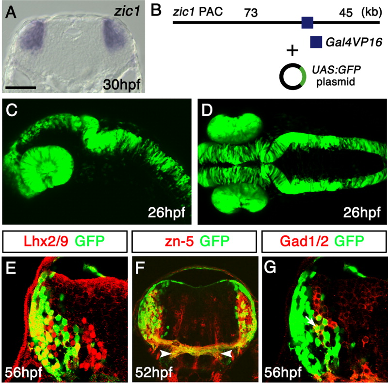
Fig. 3 Green fluorescent protein (GFP) expression in the Tg(zic1:Gal4VP16/UAS:GFP) embryos. A: In situ hybridization of a 30-hours postfertilization (hpf) coronal section of the hindbrain at the level of the otic vesicle, using the zic1 probe. Dorsal is to the top. B: The structure of the DNA constructs used to generate the Tg(zic1:Gal4VP16/UAS:GFP) fish. The zic1 PAC with Gal4VP16 inserted into the 5′-untranslater region (5′-UTR) of zic1 was coinjected with the UAS:GFP plasmid. C,D: Lateral (C) and dorsal (D) views of the Tg(zic1:Gal4VP16/UAS:GFP) embryos at 26 hpf. GFP is expressed in the eyes and dorsal neural tube. E-G: Cross-sections of the hindbrain of the Tg(zic1:Gal4VP16/UAS:GFP) embryos stained with the Lhx2/9 (E), zn-5 (F), and Gad1/2 (G) antibodies. The lateral GFP(+) cells predominantly coexpressed Lhx2/9 (E) and the zn-5 antigen (F), while some of the medial GFP(+) cells coexpressed Gad1/2 (G). A representative double-positive cell is indicated with an arrow in G. (F) The axons of the GFP(+) neurons colocalized with the zn-5(+) commissural axons. These axons projected ventrolaterally after crossing the midline (arrowheads). They were connected with the longitudinal fascicles as shown in Figure 4B. UAS, upstream activating sequences. Scale bars = 60 μm in A, 100 μm in C,D, 25 μm in E,G, 50 μm in F.
Image
Figure Caption
Figure Data
Acknowledgments
This image is the copyrighted work of the attributed author or publisher, and
ZFIN has permission only to display this image to its users.
Additional permissions should be obtained from the applicable author or publisher of the image.
Full text @ Dev. Dyn.

